Cancer Imaging
Nanomedicine holds great promise to improve the diagnosis and therapy monitoring of cancer in pediatric patients. Our team is interested in the development and clinical translation of diagnostic and combined diagnostic and therapeutic (theranostic) iron oxide nanoparticles for the benefit of children with cancer. Over the past 15 years, we have worked with every superparamagnetic iron oxide nanoparticle compound that was translated to clinical applications. We have developed a number of novel imaging applications with the FDA-approved nanoparticle compound ferumoxytol: We discovered a new approach for MR imaging of tumor associated macrophages (TAM) in mouse models (Clin Ca Res 2010) and carried out the first clinical trial on TAM imaging in pediatric cancer patients (ongoing). This approach will be used in the future to monitor new TAM-directed cancer immunotherapies. We conjugated ferumoxytol nanoparticles to an enzyme-activatable cytotoxic drug, thereby creating highly specific cancer therapeutics without side effects (Small 2014 and MCT 2017). In the process, we discovered unexpected intrinsic therapeutic effects of nanoparticles (Nature Nanotechnology 2016). We introduced novel radiation-free whole body MR imaging tests for pediatric oncology patients (Lancet Oncology 2014) and integrated this technique into PET/MR technologies for "one stop" cancer staging. We are currently translating these imaging technologies to routine clinical services. We also developed novel imaging tests for detection of chemotherapy-induced tissue injuries in pediatric cancer survivors before clinical symptoms occur. We have formed a consortium with seven Pediatric Children's Hospitals to work together on the clinical implementation of the most advanced imaging technologies currently available for the benefit of children with cancer.
Cancer Imaging
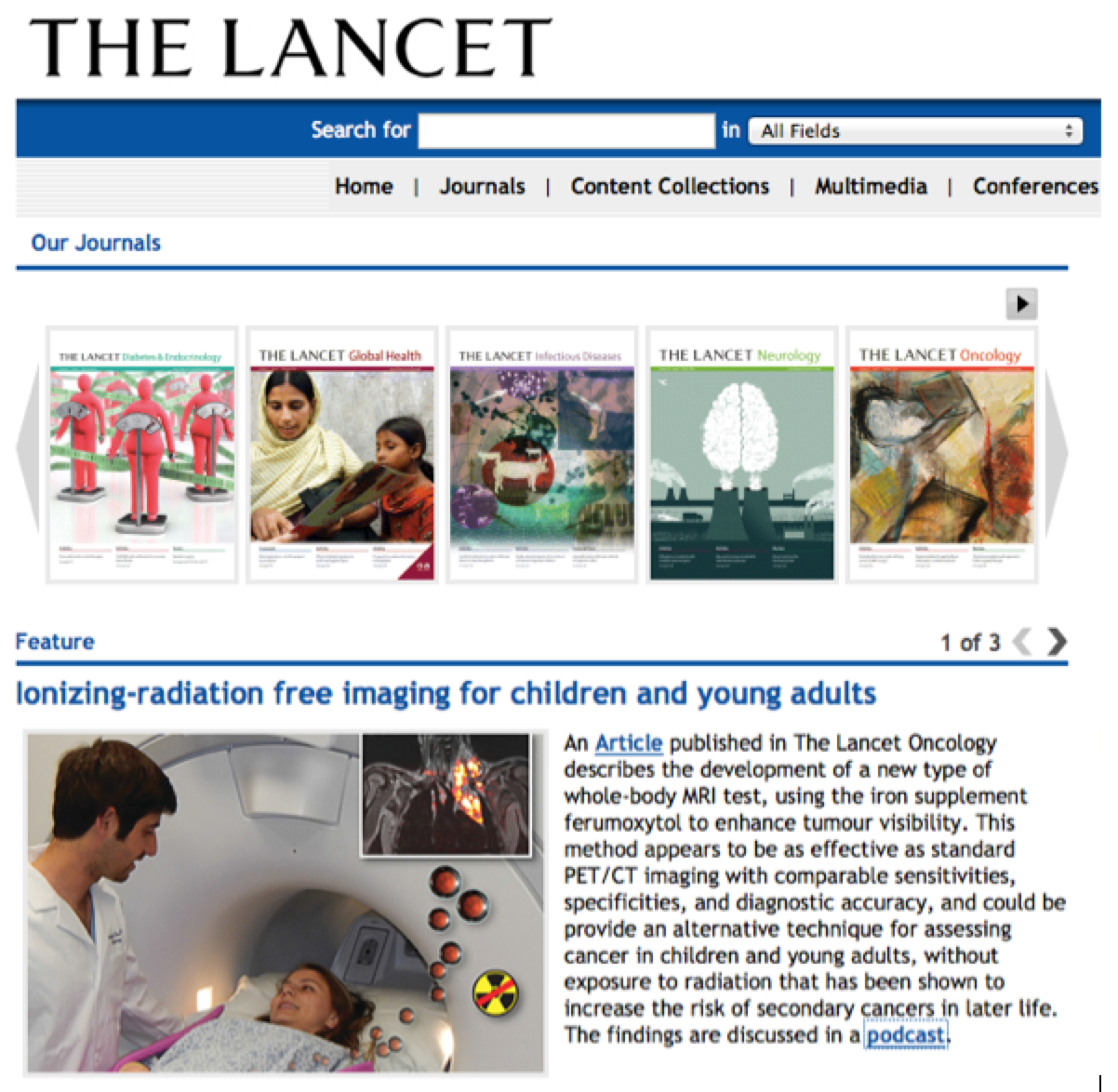 Daldrup-Link lab research featured on the website of 'The Lancet'
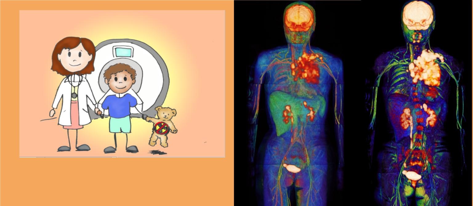
Whole Body Tumor Staging
Radiographic staging tests are essential for the diagnosis of childhood tumors, because the extent of the primary tumor and the number and location of metastases determine appropriate therapies. However, recent studies have shown that the radiation exposure associated with radiographic imaging tests may increase the risk of developing secondary malignancies later in life. This risk is particularly concerning for children, because pediatric patients are more sensitive to radiation than adult patients and they life long enough to encounter secondary cancers. We are developing novel whole body diffusion-weighted MR imaging technologies as a radiation-free alternative to standard PET/CT staging procedures. We add cell-specific contrast agents to improve the sensitivity and specificity of these new whole body staging tests. This new radiation-free imaging test may solve the conundrum of long-term side effects from radiographic staging procedures.
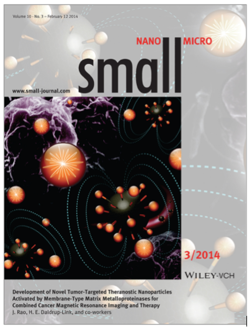 Cover article: Small 10(3): 417
Developing Cancer Therapy without Side Effects
The efficacy of traditional cancer chemotherapies is hindered by undesired dose-limiting activity upon non-cancerous organs and a lack of tools for in vivo monitoring of drug biodistributions. Approaches to improving therapeutic efficacy and monitoring whilst simultaneously reducing dose-limiting toxicities are critically needed. A new, emerging strategy is to deliver vascular-disrupting therapeutic agents (VDAs), which easily reach their target endothelial cells, and trap and dose intensify therapeutics at the tumor site. In order to avoid concomitant toxic effects in normal organs, nontoxic VDA-prodrugs can be designed, which are activated by specific enzymes in the tumor microenvironment. We integrated the concepts of MMP-14 (matrix metallo-proteinase) activatable VDA prodrugs with iron oxide nanocarrier platforms, which allows for in vivo drug tracking with MR imaging and tumor delivery of a significantly increased drug payload. We are studiing the diagnostic and therapeutic properties of novel MMP-14 activatable theranostic nanoparticles (TNPs), which exert selective vascular disruption and toxic effects in MMP-14 expressing cancers, but not normal organs, and enable real-time monitoring of drug accumulation in tumors with MR imaging.
 Cover article: Clinical Cancer Research 17(17):5695
Imaging Tumor Associated Macrophages
The presence of tumor associated macrophages (TAM) in cancer correlates strongly with tumor progression and poor outcome. While the degree of TAM infiltration has been correlated with tumor aggressiveness and overall survival in a wide range of malignant tumors, no diagnostic tool currently exists which could assess TAM quantities in a given patient's tumor non-invasively and repetitively. We are developing immediately clinically applicable, non-invasive imaging assays for selective targeting and visualization of TAM in malignant lymphomas, sarcomas, adenocarcinomas and glioblastomas. The approach is based on "off label" use of the FDA-approved iron supplement ferumoxytol as a contrast agent for MR imaging. Ferumoxytol is composed of iron oxide nanoparticles, which are phagocytosed by TAM and which can be detected with MR imaging. The new TAM imaging test can be used as a novel prognostic assay for stratifying individual patients to personalized therapies and to monitor response to novel immunotherapies that are currently entering clinical practice.
Improving Drug Delivery to Tumors
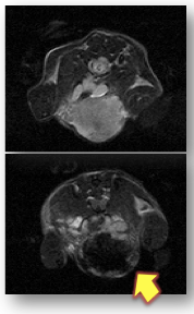
Advances in our understanding of cellular, molecular and genomic characteristics of malignant tumors has led to the development of a wide range of cell-targeted imaging probes, which are designed to provide more sensitive and specific information about the underlying tumor than conventional, non-specific contrast agents. However, one major bottleneck for successful clinical translation of these new imaging probes is their large size, which limits their delivery to tumor cells or other target cells in the tumor microenvironment. Thus, these probes currently only exert a fraction of their maximal potential. Two major barriers for efficient delivery of macromolecular drugs to the tumor interstitium include limited permeability of tumor microvessels to macromolecules and high interstitial fluid pressure (IFP) in cancers. We are developing and testing novel drug strategies that decrease IFP and increase tumor microvascular permeability and imaging enhancement of diagnostic and therapeutic macromolecular drugs (Figure shows delivery of superparamagnetic iron oxide nanoparticles to a MMTV PyMT adenocarcinoma (arrow), as demonstrated by strong negative (dark) tumor enhancement on a T2-weighted MR scan).
Clinical Trials
Pilot Development of Radiation Free Whole Body Magnetic Resonance (MR) Imaging Technique for Staging Children With Cancer
A research study on the diagnosis of spread of disease for children who have been diagnosed with solid tumors using a new whole body imaging technique and a new MR contrast agent (ferumoxytol). Standard tests that are used to determine the extent and possible spread of a child's disease include magnetic resonance (MR) imaging, computed tomography (CT), Positron Emission Tomography (PET) as well as bone scanning, and metaiodobenzylguanidine (MIBG) scanning. The purpose of this study is to determine if newer imaging tests referred to as whole body diffusion-weighted MR and whole body PET/MR can detect the extent and spread of the disease as accurately or even better as the standard tests (CT, MR and/or PET/CT). The advantage of the new imaging test is that it is associated with no or significantly reduced radiation exposure compared to standard CT and PET/CT imaging tests. The results of whole body MR and PET/MR will be compared with that of the conventional, standard imaging studies for tumor detecting.
Differentiation of Bone Sarcomas and Osteomyelitis With Ferumoxytol-Enhanced MRI
This pilot trial studies the differentiation of bone sarcomas and osteomyelitis with ferumoxytol-enhanced magnetic resonance imaging (MRI). Imaging procedures that allow doctors to more accurately differentiate between malignant bone sarcomas and osteomyelitis may help in diagnosing patients correctly and may result in more timely treatment.